ACL
Anterior cruciate ligament stops the shin bone (tibia) from sliding forward under the thigh bone (femur).
There are several ways in which the ACL can get injured.
- Rapid change of direction
- Reducing pace from an activity such as running
- Landing hard from a high jump
- Direct contact like in a football/rugby tackle
WHAT THE ACL IS
Anterior cruciate ligament stops the shin bone (tibia) from sliding forward under the thigh bone (femur).
There are several ways in which the ACL can get injured.
- Rapid change of direction
- Reducing pace from an activity such as running
- Landing hard from a high jump
- Direct contact like in a football/rugby tackle
DIAGNOSING ACL INJURY
To diagnose ACL injury, a careful history is taken to identify the mechanism of injury. Usually several clues in the history are indicative of an ACL injury and this can then be confirmed with a thorough knee physical examination. Magnetic Resonance Imaging (MRI) is used to confirm a torn ACL. Occasionally this may be confirmed with an arthroscopic inspection.
There may be a partial or a complete ACL tear. Partial tears are not often as serious as complete tears and surgical treatment may not be necessary. Complete tears may however require reconstruction.
IMMEDIATE TREATMENT
To minimise swelling, bleeding and avert further injury, you should rest the knee immediately after sustaining the injury.
Lightly tie an ice pack onto the injured knee, (best applied around 10-15 minutes two times an hour, several times throughout the day). The ice contracts the blood vessels around the affected area, thereby reducing swelling. It also helps in minimising muscle spasms and pain by numbing the nerve endings.
It is not often advisable to apply creams, massage or heat to a knee that is intensely swollen as it raises blood supply which worsens the pains and swelling.
Normally, after a couple of weeks, the pain should subside. However, it is likely for the knee to feel unstable, like it’s going to crumble when you put weight on the affected leg. Physiotherapy, braces and in certain cases, surgery is used to treat the instability.
There are certain cases in which other knee parts are injured alongside the ACL, the cartilage/meniscus particularly.
TORN ACL TREATMENT
Treatment for torn ACL can either be conservative or operative, i.e. non-surgical treatment and surgical treatment.
NON-SURGICAL TREATMENT
Non-surgical treatment may be implemented using intensive physiotherapy. This involves a treatment program called the ACL rehabilitation program which is an intensive physiotherapy to strengthen the knee and to counteract any potential instability.
ACL SURGERY
There are three primary objectives of ACL surgery and rehabilitation. They are:
1. Restoring the normal anatomy of joints
2. Providing both dynamic and static knee stability
3. Proceeding with normal life such as sports and work as soon as possible.
Often you will be sent for “Pre-habilitation” which is designed to optimize the knee prior to a surgical procedure.Your active participation in the rehabilitation prior to and after the procedure is crucial. Given the high failure rate for direct repair of the torn ACL, a reconstruction procedure is carried out using a ligament graft.
There are various options for ACL graft. These include use of the hamstring tendons, patellar tendons or use of allograft taken from a donor.
A typical ACL reconstruction involves drilling a diagonal tunnel through the thigh and shin bones. A new ACL is then inserted into the tunnel and small screws and/or buttons are used to hold it into place.
GRAFT TYPES
PATELLAR TENDON
This is the tendon that links the patella/kneecap to the tibia/shin. A portion of this is taken to act as an artificial ACL.
THE HAMSTRING TENDON
The hamstring tendons are taken and stitched together to develop a strong artificial ACL.
AN ALLOGRAFT
The allograft is the third option. It is the tissue acquired from a donor. Its main advantage is the fact that the surgeon does not have to take ligaments from elsewhere in the knee.
POST-SURGERY PHYSIOTHERAPY
A rehabilitation program should be commenced immediately by your physiotherapist to counter the swelling and pain that follows the surgery.
Surgeons and physiotherapists are constantly seeking a better understanding of the best ways to rehabilitate the knee after an ACL reconstruction. Based on the extensive research, rehabilitation programs have been developed to optimise and accelerate rehabilitation programs to allow safe, effective rehabilitation. This is effective as it stops the knee from growing stiff.
There are a number of symptoms that you will experience after the surgery which slowly fade away as time goes by. Lower leg swellings, numbness and bruising around the scars are among these symptoms but they usually disappear with time.
REHABILITATION PROGRAM
Then rehabilitation program is generally organised into the following steps:
STAGE 1: FIRST TWO WEEKS POST-SURGERY
The aim of the first stage is to minimise swelling, recover the leg’s muscle control, restore the normal posture and recover the ability to flex and extend the affected knee.
The physiotherapist is keen on getting the patient to assume an upright posture and bearing weight with a little support. They also aim at getting the patient to walk without a limp. Physiotherapists may use compression and ice to curb selling and recommend regular but short bursts of exercise.
Reduction of knee swelling depends, to a certain degree, on the ability to extend and lock the knee fast. This is because it facilitates the pumping of blood from the knee by the quadriceps muscle.
Calf and hamstring stretches, gentle bending and static quadriceps contractions are some of the exercise in this stage.
STAGE 2: TWO TO SIX WEEKS POST-SURGERY
The aim here is to eliminate use of crutches, strengthen knee, restore complete flexion and regain confidence in the knee.
Exercises involved are static cycling, climbing stairs, knee lunges and bends. Introduction of weights is done in this stage and swimming is allowed as long as it not breast stroke unless cleared for it.
STAGE 3: SIX TO TWELVE WEEKS POST-SURGERY
The aim in this stage of the program is to boost balance and coordination. Speed and resistance training is increased at this stage. Strengthening of the quadriceps and hamstring will also be continued.
STAGE 4: THREE TO SIX MONTHS POST-SURGERY
The aim in this stage is to maintain the motivation, boost aerobic fitness and develop particular sport skills.
Activities included in the program are agility skills, running and custom sports program. Others such as rotating and jumping may be introduced at this stage.
STAGE 5: OVER SIX MONTHS
This stage of the program is aimed at getting the patient back to normal level of activity. A training of three to four months may be necessary to be back to the normal level of activity in which they may be able to compete at sports. This stage is planned by the physiotherapist based on the progress of the patient.
POTENTIAL COMPLICATIONS
In most cases, the procedure is successful. However, there are a number of risks that may be involved. Some of them are;
ANAESTHETIC
– The anaesthetist will go through this with you in more detail.
INFECTION
A small risk of joint infection after the procedure exists. To prevent infections, antibiotics are usually administered during the operation. Some of the symptoms of infection include pain, swelling, redness on the wound, and even fever. Contact your doctor as soon as you detect these. There is also a small risk of chest and water work infections.
DEEP VEIN THROMBOSIS/ PULMONARY EMBOLUS
Acute pain on the calf is often a sign of blood clot in the vein. Shortness of breath/ chest pain are typical symptoms of a pulmonary embolus. Support stocking and medication to prevent these symptoms are given following the operation and need to be taken for 2 weeks post-surgery.
FAILURE
There is the risk of the knee continuing to be unstable or instability developing over time.
STIFFNESS
NEURO-VASCULAR INJURY
Damage to blood vessels or nerves can also occasionally occur following knee replacement surgery. This is rare complication but something you need to be aware of.
Related Articles
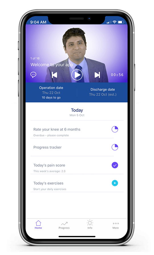
Surgery Preparation & Recovery app
Mr. Atif Malik has created an app to help his patients get ready for surgery and reach their treatment goals.
Click here to register with Mr. Atif Malik’s app