Knee Arthroscopy
ARTHROSCOPY DEFINED
Commonly referred to as Keyhole surgery. This allows an accurate assessment of the knee joint with the ability to undertake therapeutic procedures. It involves insertion of a camera into the knee and visualising the inside of the knee on monitor.
Two small incisions, usually less than a 0.5cm each are utilised. One incision is used to insert the camera whereas the other incision is used to insert specialised instruments which allows various procedures to be carried out. This include repairing or removing torn meniscal cartilage, reattaching or removing loose fragments of articular cartilage or cleaning out tissue fluid or debris.
REASONS FOR ARTHROSCOPY
It may be conducted for diagnostic purposes, such as investigating swelling, pain or joint instability whereas other investigative modalities may not have provided clear information.
Damage to ligaments or cartilage within a joint can be visualised during an arthroscopy together with providing an assessment of arthritis, loose bodies and inflammation.
Two semi-circular soft fibrocartilage structures known as the menisci which act as joint cushions often suffer injuries from twisting activities. For young adults or children, it is often possible to repair torn cartilage by trimming the affected area beck to normal.
During pivoting and twisting activities, the knee is offered stability by cruciate ligaments, (anterior and posterior). They may be injured and can be viewed clearly and physically examined through arthroscopy. If articular cartilage damage is evident then a procedure called Micro-fracture is one of the ways of treating the surface of the cartilage. This can also be carried out during a arthroscopy.
PRIOR TO ARTHROSCOPY
The knee may often be painful and swollen prior to an arthroscopy. Some of the preparation prior to the arthroscopy include discussions with the physiotherapist regarding:
- resuming work
- use of crutches
- driving
- maintaining activity
- Post-operative rehabilitation
THE ACTUAL PROCEDURE
Typically, arthroscopies last between 20 and 60 minutes, depending on the procedure. You are admitted to the hospital for the surgery on the day and are often put under a general anaesthetic. Certain occasions may call for an overnight stay.
WHAT NEXT AFTER ARTHROSCOPY
After the procedure, you are instructed on when to commence physiotherapy and how much weight to put on the leg. A summary of the operation is sent to the physiotherapist and GP.
The follow-up are arranged in the outpatient environment usually 2-6 weeks after the procedure.
POTENTIAL COMPLICATIONS
Infection:
THROMBOSIS:
Risk of thrombosis is also raised by the use of oral contraceptive pills and HRT. They should therefore be discontinued before the surgery.
BLEEDING:
this may happen after the procedure and may require the patient to go for blood and blood clot removal especially when severe.
FAILURE TO IMPROVE
NEURO-VASCULAR INJURY:
Though rare, structures near or inside the joints may be injured during the procedure.
AFTER-SURGERY CARE FOR THE KNEE
Elevate the knee and ice it to minimise swelling and reduce both inflammation and pain. Icing should be done for 15-20 minutes throughout the day between intervals of about 1-2 hours.
It is often advisable to take anti-inflammatories or pain killers for the first 5 or so days even if you do not experience pain.
There are certain symptoms that you may need to contact the doctor if you observe. They include the following:
- Bleeding on the affected area after the first two days
- Swelling that is not remedied by elevation or icing
- Tingling sensation and numbness and colour or temperature changes on the affected leg
- Stomach upset or nausea from anti-inflammatories
Related Articles
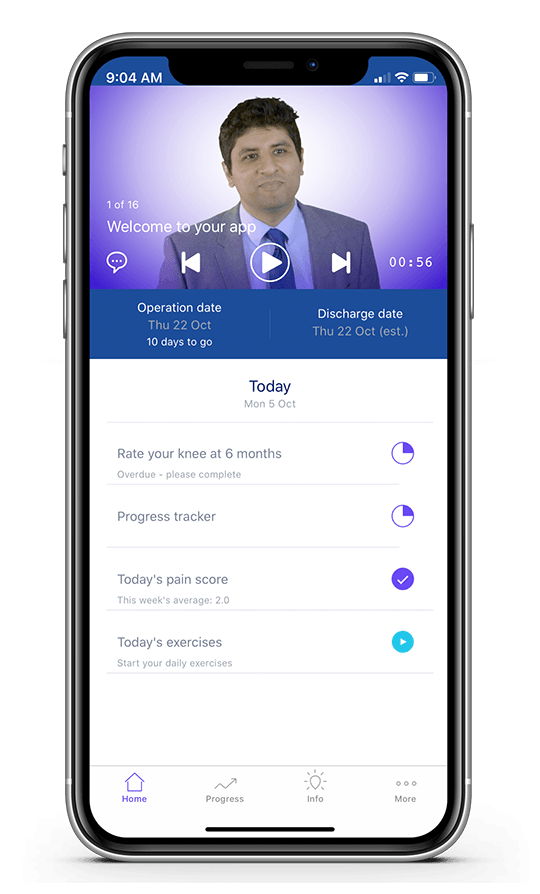
Surgery Preparation & Recovery app
Mr. Atif Malik has created an app to help his patients get ready for surgery and reach their treatment goals.
Click here to register with Mr. Atif Malik’s app